Spontaneously dislocated lens with ring-shaped pigmentation
Article Sidebar
Published:
Jun 27, 2026
Main Article Content
Abstract
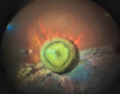
A 78-year-old man presented at Aravind Eye Hospital, Puducherry, with a 2-year history of poor vision in the left eye. He had no history of trauma or cataract surgery. On examination, best-corrected visual acuity in the left eye was 2/60. Slit-lamp examination showed a nondilating pupil measuring 2 mm diameter, with aphakia in the pupillary plane. B-scan ultrasound was suggestive of a spontaneously dislocated lens. During pars plana vitrectomy, spontaneously reattached retina was noted, with atrophy, and the lens with a near mature nucleus, outlined by a pigmentary ring, was located at the demarcation line.
Downloads
Download data is not yet available.
Article Details
How to Cite
1.
Dave P, Sonawane NJ. Spontaneously dislocated lens with ring-shaped pigmentation. Digit J Ophthalmol. 2026;32(2). Accessed July 22, 2026. https://djo.harvard.edu/index.php/djo/article/view/1410
Issue
Section
Images & Videos

This work is licensed under a Creative Commons Attribution-NonCommercial-NoDerivatives 4.0 International License.