Macular telangiectasia type 2 complicated with choroidal neovascularization
Article Sidebar
Main Article Content
Abstract
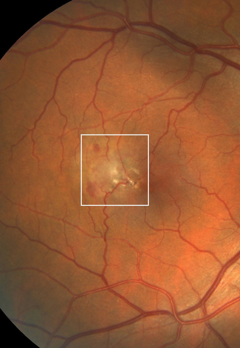
A 50-year-old man presented at Ulucanlar Eye Training and Research Hospital with decreased vision in his right eye. On examination, his best-corrected visual acuity was 20/70 in the right eye and 20/25 in the left eye. Fundus examination revealed parafoveal loss of transparency, right angle venules, and telangiectatic vessels, more prominent temporally in the macula of each eye, in addition to a dirty, grayish retinal neovascular membrane with retinal hemorrhages temporal to the fovea of the right eye. There were also retinal pigment epithelium (RPE) atrophic changes, RPE hyperplasia, and yellowish crystalline deposits in the macula of the left eye. Fundus fluorescein angiography of the right eye showed staining of the temporal parafoveal capillaries and presence of juxtafoveal choroidal neovascularization. Optical coherence tomography angiography of the right eye revealed the telangiectatic vessels in the superficial and deep retinal plexus, neovascular membrane composed of loops, peripheral anastomoses, surrounded by a hypointense halo in the outer retina and choriocapillaris slabs in addition to subretinal hyperreflective material with intraretinal fluid on the B-scan.
Downloads
Article Details

This work is licensed under a Creative Commons Attribution-NonCommercial-NoDerivatives 4.0 International License.