Focal scleral nodule
Main Article Content
Abstract
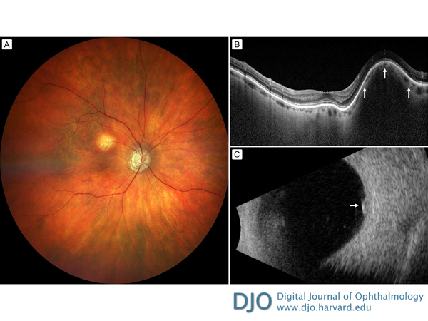
A 91-year-old white woman with a history of glaucoma presented at Wills Eye Hospital with blurred vision in the right eye and visual acuity of 20/25. On fundus examination of the right eye (A), there was a deep, yellow mass with overlying retinal pigment epithelial loss, measuring 2 mm in basal diameter and 2 mm in thickness, and located 1 mm superior to the foveola. Optical coherence tomography localized the mass within the sclera as a focal scleral nodule, with draping and slight thinning of the overlying choroid (B, arrows). Ultrasonography showed a hyperechoic, dense mass arising from the sclera and confirmed the thickness to be 2 mm (C). Observation was advised, and 6-month follow-up showed the findings to be stable. These lesions may appear similar to malignant ocular tumors; however, they arise from the sclera and remain unchanged.
Downloads
Article Details

This work is licensed under a Creative Commons Attribution-NonCommercial-NoDerivatives 4.0 International License.
