A quadruple fundal coloboma
Article Sidebar
Main Article Content
Abstract
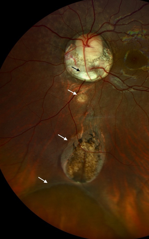
An 18-year-old girl presented at Guru Nanak Eye Center with a complaint of blurred vision in her left eye since childhood. On examination, her eye was found to have a complete typical iris coloboma (A) with visible inferior lens equator and absent zonules (B). Fundus examination revealed a normal optic disc surrounded by peripapillary atrophy enclosing a chorioretinal coloboma (C, black arrow) at its inferior border and a combination of Ida Mann type (5, 6, 7) chorioretinal colobomas (C, white arrows) connected by bridging retinal tissue. The occurrence of fundal coloboma is postulated to be due to failure of posterior closure of embryonic fissure in the optic cup. This defect usually gives rise to an isolated fundal coloboma and very rarely to multiple discrete fundal colobomas as shown here.
Downloads
Article Details

This work is licensed under a Creative Commons Attribution-NonCommercial-NoDerivatives 4.0 International License.