Posterior embryotoxon
Article Sidebar
Main Article Content
Abstract
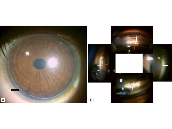
A 6-year-old boy presented at All India Institute of Medical Sciences, Gorakhpur, for evaluation of low vision in his right eye. On examination, he had best-corrected visual acuity of 20/200 in the right eye and 20/20 in the left eye, with a prescription of +6.00 +0.50 ×120 and +2.00 +1.00 ×60, respectively. Slit lamp examination revealed bilateral 360° posterior embryotoxon (A, arrow), an anteriorly displaced Schwalbe’s line, which is visible as a thin gray-white arcuate ridge on the posterior surface of the cornea. Gonioscopy showed the prominent Schwalbe’s line (B, white arrow), with insertion of pectinate strands from the iris to this line (red arrow). Fundus examination, intraocular pressure, and systemic examination were normal. He was diagnosed with anisometropic amblyopia with posterior embryotoxon and was started on occlusion therapy. Posterior embryotoxon may occur as an isolated finding or in presence of other ocular anomalies, including Axenfeld-Riegers syndrome or systemic disease, such as Alagille syndrome.
Downloads
Article Details

This work is licensed under a Creative Commons Attribution-NonCommercial-NoDerivatives 4.0 International License.