The colossal choroid
Article Sidebar
Main Article Content
Abstract
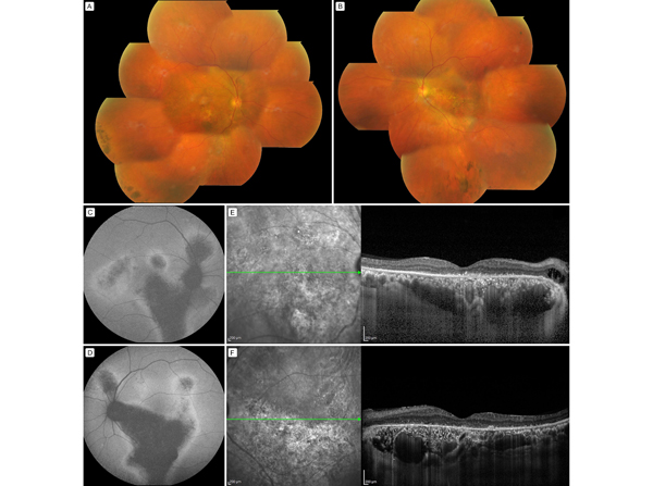
A 59-year-old man was diagnosed at Vitreoretina Services, Aravind Eye Hospital and Postgraduate Institute of Ophthalmology, Pondicherry, with chronic central serous retinopathy (CSC). Fundus examination revealed pigmentary tracts at the posterior pole in each eye, with fluid tracts and pigmentary changes (A-B). Autofluorescence imaging showed these tracts as hypofluorescent, with hyperfluorescent borders (C-D). Enhanced-depth imaging optical coherence tomography demonstrated loss of the ellipsoid zone and disrupted external limiting membrane, pachychoroid (defined as a choroidal thickness of >390 μm), and large, patchy vessels, with subfoveal choroidal thickness of 534 µm in the right eye and 396 µm in the left eye (E-F). CSC is on the pachychoroid disease spectrum. Elderly patients tend to suffer greater damage to the choroid, with retinal thinning and worse visual outcome.
Downloads
Article Details

This work is licensed under a Creative Commons Attribution-NonCommercial-NoDerivatives 4.0 International License.