Fundus photography in nonaccidental trauma
Article Sidebar
Main Article Content
Abstract
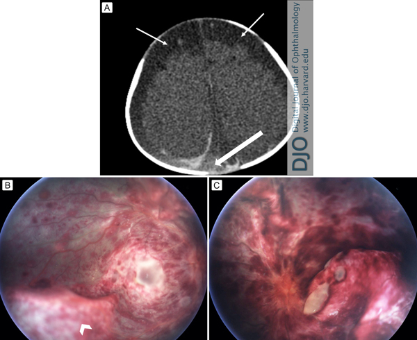
A 2-month-old previously healthy boy was presented emergently at William Beaumont Hospital, Royal Oak, Michigan, unresponsive with pulseless electrical activity and requiring cardiopulmonary resuscitation (CPR). Computed tomography of the head (A) showed acute (thick arrow) and chronic subdural (thin arrow) hemorrhages, with loss of gray white matter differentiation. On examination, pupils were fixed and unreactive to light. Fundus examination of the right eye (B) and the left eye (C) demonstrated extensive sub- and intraretinal hemorrhages throughout the retina, optic nerve edema, and multiple focal areas of retinoschisis (arrowheads). The severity of these findings suggested abusive head trauma. Other broad considerations as the cause of the ocular findings included CPR, sepsis, trauma related to vaginal delivery, and blood dyscrasias. A systemic workup was completed, including complete blood count, metabolic panel, and markers for hematologic abnormalities, which revealed no significant abnormalities. Over the next day, the patient’s systemic condition deteriorated rapidly, and he died shortly after presentation. This case highlights the importance of expedited ophthalmic consultation and photodocumention using a portable fundus camera where abusive head trauma is suspected.
Downloads
Article Details

This work is licensed under a Creative Commons Attribution-NonCommercial-NoDerivatives 4.0 International License.