Wyburn-Mason syndrome
Article Sidebar
Main Article Content
Abstract
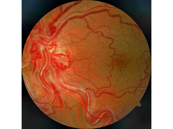
A 40-year-old man presented at Clinica Vision Total with decreased vision in his left eye. He had a history of vitreous hemorrhage and retinal detachment in his right eye, without early surgical management. Past medical history included surgery for an arteriovenous malformation at the level of the brainstem, without neurological sequelae. On initial ophthalmologic examination, his best-corrected visual acuity was no light perception in the right eye and 20/200 in the left eye. The right eye was phthisical. Anterior segment examination of the left eye was normal. Fundus examination revealed retinal arteriovenous malformations (AVMs), with markedly convoluted, dilated, and tortuous retinal vessels extending from the disc. Fluorescein angiography demonstrated rapid filling of the vascular anomalies, without leakage. The patient is followed by a multidisciplinary cohort of physicians and managed conservatively. Wyburn-Mason syndrome is a rare, nonhereditary disorder in which vascular dysgenesis affects the retina and brain. It is characterized by the presence of AVMs that vary in size and location. These lesions are direct artery-to-vein communications without an intervening capillary system to mitigate the high-flow arterial blood. There are no known genetic causes or risk factors associated with the disease. The visual acuity of patients with AVMs of the retina can be normal to mildly reduced when AVMs occur locally, in contrast to more extensive AVMs, which cause significantly reduced vision. Management is generally conservative, although surgical options are available. Regular ophthalmologic evaluations are required to monitor AVMs and their potential complications.
Downloads
Article Details

This work is licensed under a Creative Commons Attribution-NonCommercial-NoDerivatives 4.0 International License.