Swept source optical coherence tomography imaging in macular coloboma
Article Sidebar
Main Article Content
Abstract
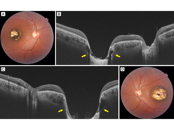
A 20-year-old man presented with outward deviation of his left eye since birth. Best-corrected visual acuity was 20/40, N12 in the right eye and 20/125, N18 in the left eye, with subnormal color vision of 8/17 in both eyes. There were no known systemic ailments. Dilated fundus examination revealed a well-defined, punched-out, macular lesion measuring about 2.5 disc diameters (DD) in right eye (A) and 4 DD in the left eye (D). The base of the lesion showed bare sclera, with several large choroidal vessels, and the edge showed retinal pigment epithelium (RPE) atrophy. These features were suggestive of bilateral macular coloboma. Swept source optical coherence tomography (Topcon DRI OCT Triton plus; Tokyo, Japan) showed deep, bowl-shaped excavations of the sclera (B, C) corresponding to the colobomatous areas, with loss of overlying neurosensory retina, RPE, and choroid. A thin, membranous structure is seen overlying the area of bared sclera, which may be the internal limiting membrane. The patient underwent strabismus surgery to correct the deviation.
Downloads
Article Details

This work is licensed under a Creative Commons Attribution-NonCommercial-NoDerivatives 4.0 International License.