Corneal deposits in nephropathic cystinosis
Article Sidebar
Main Article Content
Abstract
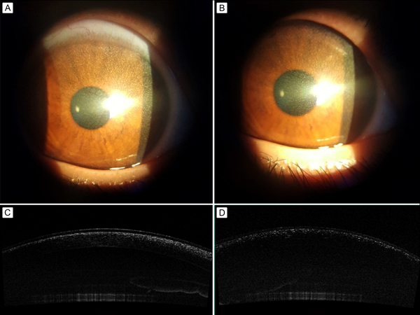
A 4-year-old girl followed in the Department of Pediatrics, in Hedi Chaker University Hospital Sfax, Tunisia, for nephropathic cystinosis, was found on ophthalmological examination to have mild photophobia. Her visual acuity was normal in both eyes. Slit-lamp biomicroscopy revealed diffuse crystal deposition throughout the entire cornea in both eyes (A-B). There was no associated corneal thinning, edema, or scarring. Intraocular pressure was normal, the anterior chamber was deep and quiet, and the lens was clear. Fundus examination was normal in both eyes. Anterior segment optical coherence tomography (AS-OCT) showed hyperreflective punctuate foci from limbus to limbus within the anterior stroma (C-D). Treatment with cysteamine eyedrops was initiated. Cystinosis is a rare disease with autosomal recessive mutation in the CTNS gene on chromosome 17p13 that can lead to intracellular accumulation of cystine crystals in the eyes, kidneys, bone marrow, liver, spleen, thyroid, pancreas, and muscles. Ocular cystinosis can manifest as crystals in any layer of the cornea. The highest density of crystals in our patient was found in anterior stroma, followed by Bowman’s layer and the middle stroma. Crystal deposits are located deeper with increasing age. In children crystals tend to locate anteriorly, whereas in adults deposits are found more posteriorly in the corneal stroma. Careful slit-lamp biomicroscopy and adjunctive AS-OCT allows determination of precise location and depth of crystals within the corneal layers.
Downloads
Article Details

This work is licensed under a Creative Commons Attribution-NonCommercial-NoDerivatives 4.0 International License.