Bull’s eye maculopathy in a young patient with systemic lupus erythematosus
Article Sidebar
Main Article Content
Abstract
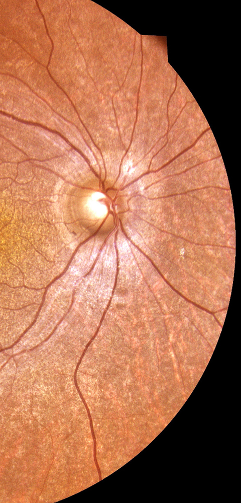
A 19-year-old woman diagnosed with systemic lupus erythematosus and treated with hydroxychloroquine therapy 200 mg/day for 6 years (cumulative dose approximately 438 g) presented at Ibn Sina University Hospital for her routine ophthalmological examination. There was no history of childhood overdose. Visual acuity was 20/40 in each eye. Funduscopic examination revealed atrophic bull’s eye maculopathy, with concentric rings of hypopigmentation and hyperpigmentation surrounding the fovea in the right eye and left eye. Optical coherence tomography showed decreased retinal thickness (foveal thickness, 191 μm), and atrophy of the outer retinal layers with interruption of the ellipsoid line. Hydroxychloroquine was discontinued after the diagnosis of retinal toxicity. At the 12-month follow-up, visual acuity remained stable, and no further progression of retinal changes was observed.
Downloads
Article Details

This work is licensed under a Creative Commons Attribution-NonCommercial-NoDerivatives 4.0 International License.