Anterior dislocated Morgagnian cataract
Article Sidebar
Main Article Content
Abstract
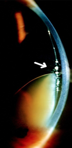
A 62-year-old man presented at the Sankara Nethralaya outpatient department with gradual, painless loss of vision in his left eye. On examination, best-corrected visual acuity in the left eye was hand movements. Right eye visual acuity was normal. He had no history of trauma or ocular surgery. Slit-lamp examination revealed anterior dislocation of a hypermature Morgagnian cataract. Anterior segment optical coherence tomography revealed a diaphanous wavy capsular bag, along with liquified cortical matter and sunken nucleus touching the corneal endothelium. A narrow slit-beam examination angled at 60° showed the “triangle sign” indicative of Morgagnian cataract—a triangular area formed by the anterior and posterior lens capsule at the apex and the lens nucleus at the base. Posterior segment ultrasonography was unremarkable. Morgagnian cataract is prone to anterior dislocation or displacement because of the degeneration of the lens suspensory ligament. Untreated Morgagnian cataract can cause capsular bag rupture, potentially resulting in phacoanaphylactic uveitis or phacolytic glaucoma. Our patient underwent successful cataractous lens removal with anterior vitrectomy and scleral fixation intraocular lens implantation with good visual recovery.
Downloads
Article Details

This work is licensed under a Creative Commons Attribution-NonCommercial-NoDerivatives 4.0 International License.