Pigmented conjunctival lesions in ochronosis
Article Sidebar
Published:
Dec 9, 2019
Main Article Content
Abstract
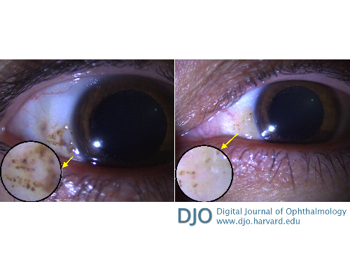
A 45-year-old man was diagnosed to have alkaptonuria after his orthopedist observed joint discoloration during knee arthroplasty. He was referred to Dokuz Eylul University, Department of Ophthalmology, for evaluation. On examination, his visual acuity was 20/20 in each eye. Slit-lamp examination revealed brownish pigmented lesions in both the nasal and temporal aspects of the sclera in the interpalpebral region (Osler’s sign) bilaterally. Fundus examination was unremarkable in both eyes.

Downloads
Download data is not yet available.
Article Details
How to Cite
1.
Saatci, MD AO, İnel, MD TY, İpek, MD Şefik C. Pigmented conjunctival lesions in ochronosis. Digit J Ophthalmol. 2019;25(4). Accessed June 17, 2026. https://djo.harvard.edu/index.php/djo/article/view/102
Issue
Section
Images & Videos

This work is licensed under a Creative Commons Attribution-NonCommercial-NoDerivatives 4.0 International License.